SUMMER 2021


A clinical study sheds new light on the efficacy of soft multifocal contact lenses used for the management of myopia


Remy Marcotte-Collard

Langis Michaud


Introduction One of the most common questions that practitioners ask us is what the best lens or strategy is to control myopia in children. Most of them do not practice orthokeratology and are looking for a single method that could be effective and user-friendly, both for patients and for themselves.

The first consideration is that there is no one-size-fits-all treatment approach that will work for everyone. Each child is different and should be treated in a personalized way. The analysis of one patient will be different from that of the next even if they are the same age, the same sex, the same ethnicity and are presenting similar clinical data, they do not have the same physiological basis and long-term risk factors.


It is in this perspective that we are interested in analyzing three commercial lenses used for the control of myopia, some off-label. Are they equally effective, or is there one that should be favored over the others? And under what circumstances? To answer these questions, there are two possible approaches. The first, classic, approach is to adapt groups of patients with one or the other of these lenses, in a randomly determined order, and to compare their evolution over 2 years with a control group equipped with glasses. This approach is scientifically rigorous but costs a lot of money and represents a heavy investment in time. We do not have quick answers, and if this approach is not conclusive, it is necessary to repeat the tests over a longer period of time with other products. We will never live long enough to do it all.


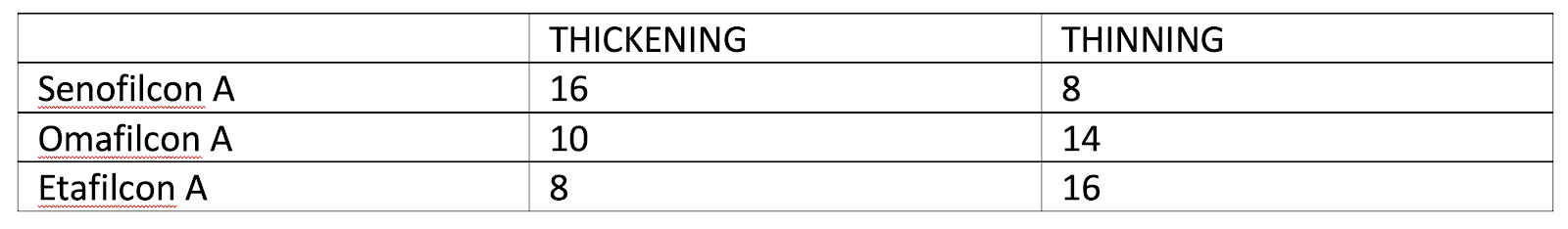
Another more pragmatic approach makes it possible to carry out short-term experiments to determine which are the most relevant elements to subsequently conduct a longitudinal study. This can save time and money while testing multiple hypotheses, without spending decades to find answers to our questions. This method is based on analyzing the variation of the choroid in response to optical stimuli. Indeed, in recent years, studies have shown that the short-term choroidal response can be considered as a valid indicator of the effectiveness of an optical device used in the management of myopia. Although there is no consensus on the technical parameters, it is agreed that a measurement with an optical coherence tomographer (OCT) allowing acquisition of quality images of the choroid is a must. Determining the choroidal thickness is then done manually or via image analysis with a given software. The use of artificial intelligence has a specific niche here. In general, when the visual stimulus involves thickening of the choroid, it is agreed that this response is considered positive and therefore promotes better control of the elongation of the eye. Conversely, when the visual stimulus leads to thinning of the choroid, it is agreed that this signal encourages the progression of myopia and axial length.


METHODS & LENSES TESTED


We recruited a cohort of 24 Caucasian young adults (12 F/12M 25.8 ± 3.5 y.o.) who had moderate myopia (-2.58D ± 1.48D). All had best-corrected visual acuity (BCVA) of 20/20 or better monocularly and binocularly, and none had been diagnosed with ocular pathology. Participants were asked to not wear contact lenses for 72 hours prior to testing. Activities and nutrition were controlled 24 hours before and on the day of the testing (refraining from athletic training or strenuous exercises, stimulating food, caffeine, alcohol, tobacco, etc.).


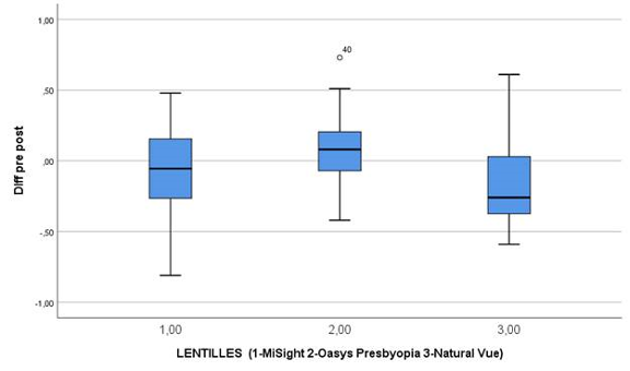
Three different soft multifocal contact lenses were selected: senofilcon A (Acuvue Oasys for Presbyopia, J&J Vision Care) omafilcon A (MiSight, CooperVision) and etafilcon A (NaturalVue, Visioneering Technology). Lenses were randomly fitted on every participant. The lenses were worn for 30 minutes followed by a 20-minute washout period. Throughout the experiment, participants had to read a book without images or watch a (boring) film that did not generate strong emotions or display much action.
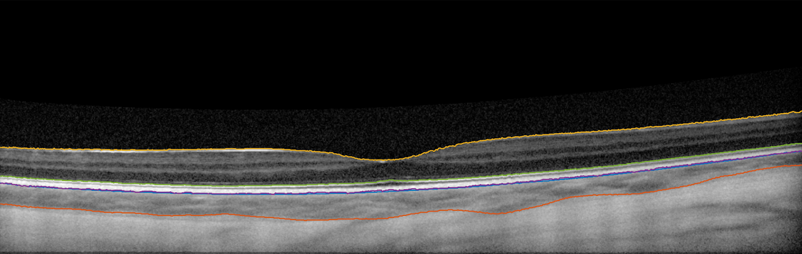
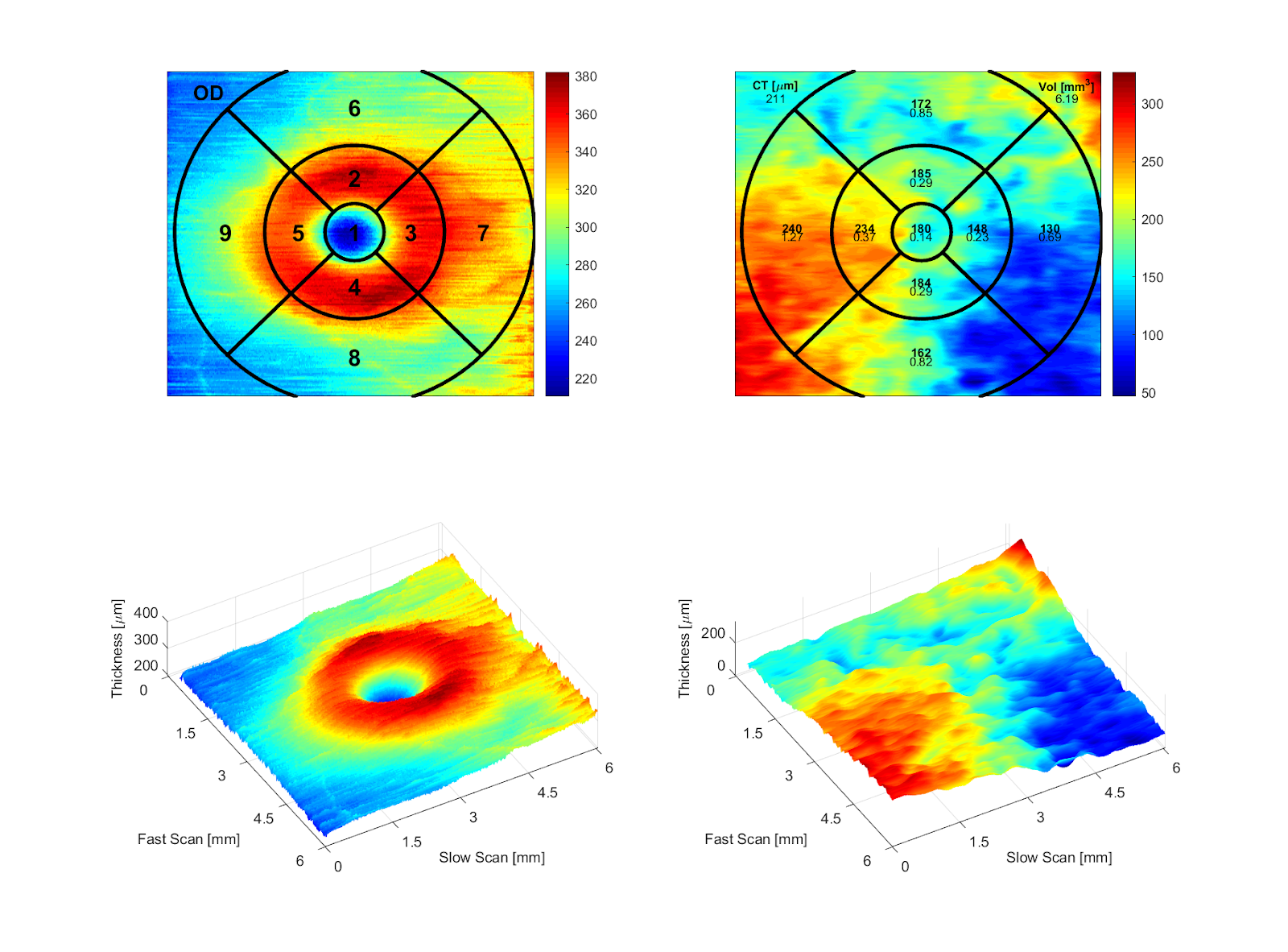
Images were taken with a confocal scanning laser ophthalmoscope (cSLO, Heidelberg Spectralis) featuring an active eye tracking and AutoRescan software, which ensures that the measured variations were based on the same locations on the retina, corresponding to the previous measurements at 100%. Images were then processed through a proprietary matlab software that was able to evaluate not only the thickness of the choroidal tissue but also its volume [1], which is more important and clinically relevant. Detection of the choroidal margins was automated with the use of artificial-intelligence-based software, following the ETDRS approach by quadrant [2].


Sample of the image taken and delineation of the choroidal margin after image processing.


RESULTS

Overall results show that the senofilcon A lens was the only one that resulted in choroidal thickening (+ 0,086 ±0,239), while the omafilcon A lens (-0,055 ±0,294) and the etafilcon A lens (-0,150 ±0,321) resulted in choroidal thinning. The only statistically significant difference was found between the senofilcon A lens and the etafilcon A lens (95% CI (0.037-0.433 p=0.015). The omafilcon A lens and the senofilcon A lens difference did not reach statistical significance (p=0.253).

As expected, there were a lot of inter-subject variations. It becomes interesting when we look at the details. The following table indicates the number of participants showing thinning and thickening of the choroid when exposed to each lens. If we go a little bit further, it is possible to report that the senofilcon A lens was the best option for 14 of the 24 participants, while the etafilcon A lens led to the best outcome for 6 participants and the omafilcon A lens for only 4 participants. Pupil diameter and refractive error was not significantly different. It is then not possible to explain the winning design by these factors.


Finally, results were also different by quadrants. Regions 3-4-5 around the macula showed an increased response vs. central or more peripheral quadrants. This is in accordance with the theory that peripheral refraction (15 degrees around the macula on each side) has more influence than does the central visual stimulus. More interesting is that the superior and temporal quadrants provide a different response compared to the nasal and inferior quadrants. This suggests a more complex retinal response than previously thought.


Colored map representing choroidal variation along ETDRS quadrants, and a second map showing volume.


DISCUSSION


This study shows that design matters in myopia management, because different lenses led to a different choroidal response and in different quadrants. It is important to carefully interpret these results, as the same design does not seem to work for everyone. The senofilcon A lens was the best option for most participants (14/24), but 10 were better adapted with other designs. In fact, 6 were better served with the etafilcon A lens, while only 4 showed better results with the omafilcon A lens. For 2 participants, all lenses led to choroidal thinning, suggesting that none of them would had been effective over the long term to manage myopia or eye elongation.


It is important to note that we talk here about an increase or a decrease, in percentage, of the choroidal volume after short-term exposure to a visual stimulus through the tested devices. This was found predictive of long-term results, but more work needs to be done to extrapolate their impact in the future. This does not translate to refractive change or axial length modification while wearing lenses. Results are predictive, then non-conclusive.


It is possible to understand these results in light of a threshold hypothesis formulated by Earl Smith. In fact, he showed that visual signals entering the eye are managed by 2 different neurological pathways, one for the myopic defocus and the second responding to the hyperopic defocus. Visual stimulation generates a response at a threshold level that is different for each individual. This may explain why some participants were responding more to one design than to another: one design helps to reach the individual threshold at which the myopic defocus overcomes the hyperopic defocus, leading to choroidal thickening.


Design impact is based on the central and peripheral powers, knowing that there is a dose response, the specific areas of each of these powers, the transition between them, etc. It is then difficult to predict which design will fit best on a particular individual. More work is needed to answer to these questions.


Another factor to consider is that the results apply only to a population similar to the tested cohort, which means young Caucasian, moderately myopic adults. Future studies would include persons of different ethnical origin and age or a population with higher refractive error.


CONCLUSION


This study reinforces the fact that myopia management strategies must be customized to each individual patient, as different devices may lead to different outcomes. Some devices that would be effective for a given patient will lead to myopia or axial length increase for another who has similar refractive error. Choroidal volume analysis is a promising new way to evaluate ocular response to visual stimulation. It may be a convenient way to test—in the short term—the individual response and to determine the best strategy for a given patient.


Acknowledgements


We would like to thank Dr. Aulne St-Amant, OD and Dr. Justine Renaud, OD for their substantial contibution in the study


References

  1. Open-source algorithm for automatic choroid segmentation of OCT volume reconstructions : Javier Mazzaferri, Luke Beaton, Gisèle Hounye, Diane N. Sayah, and Santiago Costantino, Scientific Reports 7. Article Number 42112, (2017). doi:10.1038/srep42112.
  2. Belanger Nzakimuena, C., Marcotte-Collard, R., & Simard, P. (2019). An algorithm for structuring data into an Early Treatment Diabetic Retinopathy Study (ETDRS) grid (https://github.com/cnzakimuena/ETDRSmaps), GitHub.